
Prévalence des cellules pneumatiques zygomatiques dans la population tunisienne : Etude rétrospective
DOI :
https://doi.org/10.62438/tunismed.v104i01.6051Mots-clés :
Articulation temporo-mandibulaire (ATM), Radiographie panoramique, Tomographie volumique à faisceau conique (CBCT), Études rétrospectivesRésumé
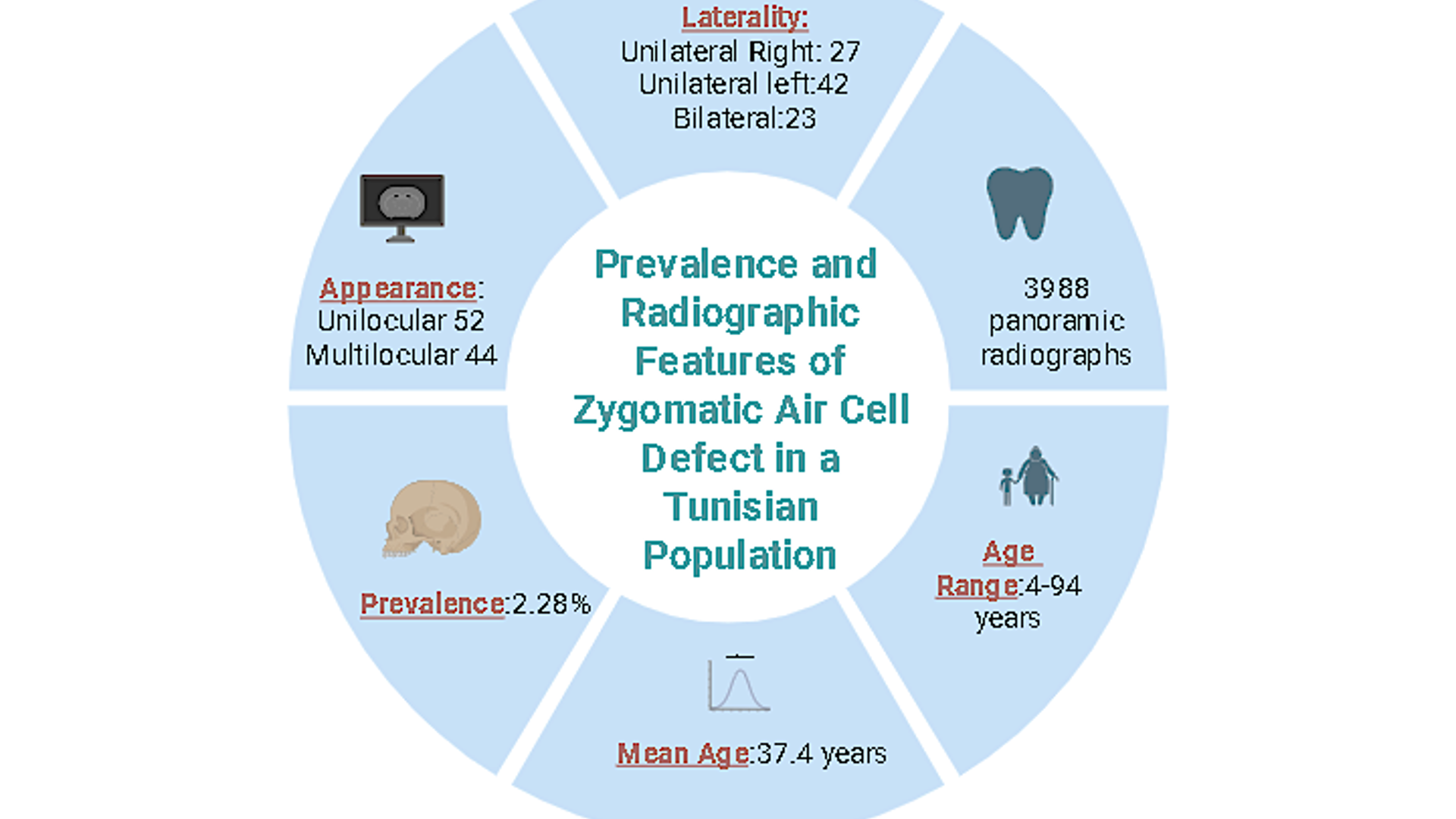
Objectifs : Les cellules pneumatiques zygomatiques (ZACDs) sont des variantes anatomiques pouvant compliquer les interventions chirurgicales impliquant l’articulation temporo-mandibulaire. Cette étude rétrospective visait à déterminer la prévalence, la localisation et les caractéristiques morphologiques des ZACDs dans une population tunisienne, et à comparer les résultats avec les données publiées.
Méthodes : Un total de 3 988 radiographies panoramiques numériques, provenant de patients âgés de 4 à 94 ans, a été évalué de manière indépendante par trois radiologues oraux et maxillo-faciaux. Les caractéristiques démographiques, la latéralité des ZACD (unilatérale/bilatérale) et la morphologie (uniloculaire/multiloculaire) ont été enregistrées. L’analyse statistique a été réalisée à l’aide du test du Chi², et les résultats ont été interprétés dans le contexte d’une revue de littérature exhaustive.
Résultats : Les ZACDs ont été détectés dans 2,28 % des cas, avec un âge moyen de 37,4 ans (extrêmes : 7–67). La majorité des cas était unilatérale (69/92), prédominant du côté gauche, tandis qu’une atteinte bilatérale a été observée chez 23 patients. Les morphologies uniloculaire (56,5 %) et multiloculaire (43,5 %) étaient presque également réparties. Aucune association significative n’a été observée entre la présence de ZACD et l’âge, le sexe, la latéralité ou le type de locularité des patients.
Conclusion : La prévalence des ZACDs dans la population tunisienne est comparable à celle d’autres populations, mais ces anomalies semblent plus fréquentes chez les sujets jeunes. La reconnaissance des ZACDs sur les radiographies panoramiques présente un intérêt clinique, car leur présence peut compliquer les procédures ....( résumé tronqué à 250 mots).
Téléchargements
Références
Shokri A, Noruzi-Gangachin M, Baharvand M, Mortazavi H. Prevalence and characteristics of pneumatized articular tubercle: First large series in Iranian people. Imaging Sci Dent.2013;43(4):283-7.
Görür K, Özcan C, Talas DÜ. The computed tomographical and tympanometrical evaluation of mastoid pneumatization and attic blockage in patients with chronic otitis media with effusion. Int J Pediatr Otorhinolaryngol.2006;70(3):481‑5.
Zamaninaser A, Rashidipoor R, Mosavat F, Ahmadi A. Prevalence of zygomatic air cell defect: Panoramic radiographic study of a selected Esfehanian population. Dent Res J. 2012;9(1):S63-68.
Tyndall DA, Matteson SR. The zygomatic air cell defect (ZACD) on panoramic radiographs. Oral Surg Oral Med Oral Pathol.1987;64(4):373‑6.
Khojastepour L, Mirbeigi S, Ezoddini F, Zeighami N. Pneumatized Articular Eminence and Assessment of Its Prevalence and Features on Panoramic Radiographs. J Dent Tehran Iran. 2015;12(4):235‑42.
Kishore M, Sunil R, Kishore A, Aggarwal A, Upadhyay N, Agarwal N. Prevalence of Zygomatic Air Cell Defect using Orthopantomogram. J Clin Diagn Res.2015;9(9):09-11
Aldhelai TA, Altawili ZM, Alsughier Z, Almashraqi AA. Prevalence of zygomatic air cell defects in a sample of 6–18-year-old patients in Saudi Arabia and Yemen: a retrospective study. BMC Oral Health. 2024;24(1):810.
Dalal DH, Smith HF. Developmental Changes in Morphology of the Middle and Posterior External Cranial Base in Modern Homo sapiens. BioMed Res Int.2015;2015:1-16.
Srikanth H, Patil K, Mahima V. Zygomatic air cell defect: A panoramic radiographic study of a south Indian population. Indian J Radiol Imaging. 2010;20(2):112‑4.
Patil K, Mahima VG, Malleshi SN, Srikanth HS. Prevalence of Zygomatic Air Cell Defect in adults—A retrospective panoramic radiographic analysis.Eur J Radiol.2012;81(5):957‑9.
Kaugars GE, Mercuri L, Laskin D. Pneumatization of the articular eminence of the temporal bone: prevalence, development, and surgical treatment. J Am Dent Assoc. 1986;113(1):55-7.
Carter LC, Haller AD, Calamel AD, Pfaffenbach AC. Zygomatic air cell defect (ZACD). Prevalence and characteristics in a dental clinic outpatient population. Dentomaxillofacial Radiol. 1999;28(2):116‑22.
Orhan K, Delilbasi C, Cebeci I, Paksoy C. Prevalence and variations of pneumatized articular eminence: A study from Turkey.Oral Surg Oral Med Oral Pathol Oral Radiol Endod. 2005;99(3):349‑54.
Yavuz MS, Aras MH, Güngör H, Büyükkurt MC. Prevalence of the pneumatized articular eminence in the temporal bone. J Cranio-Maxillofac Surg. 2009;37(3):137‑9.
Ndiaye ML, Diatta M, Gassama BC, Kane M, Bouali B, Tamba B, Lecor PA, Toure B. Pneumatisation of the temporal articular eminence: radiographic study in a Senegalese population. Mali Med. 2021;36(3):56–59.
Gupta D, Sheikh S, Pallagatti S, Aggarwal A, Goyal G, Chidanandappa RN, et al. Zygomatic air cell defect: a panoramic radiographic study of a North Indian population. J Investig Clin Dent. 2013;4(4):247‑51.
Zadravec D, Badel T, Smoljan M, Čimić S, Katavić N, Savić PI. Zygomatic Air Cell Defect - Magnetic Resonance Imaging of the Temporomandibular Joint Compared with Panoramic Radiographs. Acta Clin Croat. 2018;57(2):227-234.
Turgeon DP, Novack R. Prevalence of zygomatic air cell defects in a North American population. Gen Dent. 2023;71(1):42-46.
Gupta D, Rashmi NC, Sheikh S, Pallagatti S, Goyal G, Singh R et al. The prevalence, radiographic appearance, and characteristics of zygomatic air cell defects (ZACDs) in symptomatic temporomandibular joint disorder patients in North Indian population. Oral Maxillofac Surg. 2014;18(4):453-7.
Park YH, Lee SK, Park BH, Son HS, Choi M, Choi KS et al. Radiographic evaluation of the zygomatic air cell defect. Korean J Oral Maxillofac Radiol. 2002;32(4):207–11.
Arora KS, Kaur P, Kaur K. ZACD: A Retrograde Panoramic Analysis among Indian Population with New System of Classification. J Clin Diagn Res. 2016;10(2):71-3.
Miloglu O, Yilmaz AB, Yildirim E, Akgul HM. Pneumatization of the articular eminence on cone beam computed tomography: prevalence, characteristics and a review of the literature. Dentomaxillofacial Radiol. 2011;40(2):110‑4.
Hofmann T, Friedrich RE, Wedl JS, Schmelzle R. Zygomatic air cell defect (ZACD). Mund Kiefer GesichtsChir. 2001;5(3):173‑9.
Orhan K, Delilbasi C, Orhan A. Radiographic evaluation of pneumatized articular eminence in a group of Turkish children. Dentomaxillofacial Radiol. 2006;35(6):365‑70.
Wrzosek M, Wilczek K, Tusiewicz J, Piskórz MJ, Różyło-Kalinowska I. Pneumatization of the articular eminence in cone-beam computed tomography: prevalence and characteristics — literature review. Folia Morphol. 2023;82(3):242‑7.
Buyuk C, Gunduz K, Avsever H. Prevalence and characteristics of pneumatizations of the articular eminence and roof of the glenoid fossa on cone-beam computed tomography. Oral Radiol. 2019;35(2):171–176.
Ladeira DB, Barbosa GL, Nascimento MC, Cruz AD, Freitas DQ, Almeida SM. Prevalence and characteristics of pneumatization of the temporal bone evaluated by cone beam computed tomography. Int J Oral Maxillofac Surg. 2013;42(6):771-5.
Da Costa Ribeiro R, Dos Santos BJ, Provenzano N, De Freitas PHL. Dautrey’s procedure: an alternative for the treatment of recurrent mandibular dislocation in patients with pneumatization of the articular eminence. Int J Oral Maxillofac Surg. 2014;43(4):465‑9.
Khojastepour L, Paknahad M, Abdalipur V, Paknahad M. Prevalence and Characteristics of Articular Eminence Pneumatization: A Cone-Beam Computed Tomographic Study. J Maxillofac Oral Surg. 2018;17(3):339‑44.
ElBeshlawy D. CBCT Assessment of Pneumatization of the Articular Tubercle and the Roof of the Glenoid Fossa: a retrospective study. Egypt Dent J. 2020;66(3):1553‑62.
Fichiers supplémentaires
Publiée
Numéro
Rubrique
Catégories
Licence
Merci de créditer les auteurs lors de toute citation : La Tunisie Médicale (2026)

Ce travail est disponible sous licence Creative Commons Attribution - Pas d'Utilisation Commerciale - Pas de Modification 4.0 International.
