
Planification chirurgicale assistée par tomographie à cohérence optique du segment antérieur pour l'extraction d'un corps étranger intracornéen : Un cas clinique avec revue de la littérature
DOI :
https://doi.org/10.62438/tunismed.v102i11.5209Mots-clés :
Corps étranger oculaire, Imagerie multimodale , Lésions cornéennes, Lésions oculaires , Tomographie par cohérence optiqueRésumé
.
Introduction : Les corps étrangers cornéens représentent une urgence courante en ophtalmologie. Bien que de nombreux cas soient simples, impliquant un diagnostic et un traitement clair, certains présentent des défis significatifs en raison de la complexité du corps étranger impliqué.
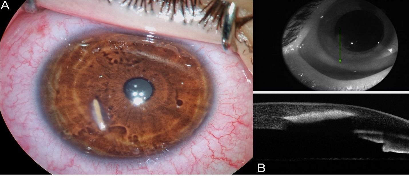
Observation : Un homme de 20 ans a présenté un inconfort dans son œil droit suite à un accident survenu en coupant un arbre sans porter de lunettes de protection. L'examen à la lampe à fente a révélé un corps étranger végétal incrusté dans la cornée. La tomographie à cohérence optique du segment antérieur a objectivé un corps étranger incrusté à 300 µm sous l'épithélium cornéen respectant les couches les plus profondes. Le corps étranger a été chirurgicalement retiré par voie antérieure sous anesthésie topique, avec suture de la porte d’entrée du corps étranger.
Conclusion : La tomographie à cohérence optique du segment antérieur est inestimable dans le diagnostic et la gestion des corps étrangers cornéens complexes, permettant une planification précise de l’approche thérapeutique, comme démontré dans ce cas.
Téléchargements
Références
McGwin G Jr, Owsley C. Incidence of emergency department–treated eye injury in the United States. Arch Ophthalmol. 2005;123(5):662–6.
Celebi ARC, Kilavuzoglu AE, Altiparmak UE, Cosar CB, Ozkiris A. The role of anterior segment optical coherence tomography in the management of an intra-corneal foreign body. SpringerPlus. 2016;5(1):1559.
Wylegala E, Dobrowolski D, Nowińska A, Tarnawska D. Anterior segment optical coherence tomography in eye injuries. Graefes Arch Clin Exp Ophthalmol. 2009;247(4):451–5.
Bhargava M, Bhambhani V, Paul RS. Anterior segment optical coherence tomography characteristics and management of a unique spectrum of foreign bodies in the cornea and anterior chamber. Indian J Ophthalmol. 2022;70(12):4284–92.
Armarnik S, Mimouni M, Goldenberg D, Segev F, Meshi A, Segal O, et al. Characterization of deeply embedded corneal foreign bodies with anterior segment optical coherence tomography. Graefes Arch Clin Exp Ophthalmol. 2019;257(6):1247–52.
Loporchio D, Mukkamala L, Gorukanti K, Zarbin M, Langer P, Bhagat N. Intraocular foreign bodies: A review. Surv Ophthalmol. 2016;61(5):582–96.
Darade DM, Naik HD. Management of an intrastromal corneal foreign body. J Clin Ophthalmol Res. 2023;11(1):57.
Volek E, Sandor GL, Nagy ZZ, Schneider M. Anterior segment optical coherence tomography-assisted surgical planning of an organic intralamellar corneal foreign body removal: a case report. Spektrum Augenheilkd. 2023; Available from: https://doi.org/10.1007/s00717-023-00551-2
Yeniad B, Beginoglu M, Ozgun C. Missed intraocular foreign body masquerading as intraocular inflammation: two cases. Int Ophtalmol. 2010;30(6):713-6.
Arora T, Arora S, Sinha R. Management of intrastromal glass foreign body based on anterior segment optical coherence tomography and Pentacam analysis. Int Ophthalmol. 2015 ;35(1):1–1.
Maeda N. Optical coherence tomography for corneal diseases. Eye Contact Lens. 2010;36(5):254.
Wang T, Zhong L, Yin S, Bao T, Yang J, Wang T, et al. Comparison of different types of corneal foreign bodies using anterior segment optical coherence tomography: a prospective observational study. J Ophthalmol. 2020; 2020:9108317.
Goel N, Pangtey BPS, Raina UK, Ghosh B. Anterior segment optical coherence tomography in intracorneal foreign body. Oman J Ophthalmol. 2012;5(2):131–2.
Akbaş E, Barut Selver Ö, Palamar M. Retrospective evaluation of corneal foreign bodies with anterior segment optical coherence tomography. Turk J Ophthalmol. 2021;51(5):265–8.
Al-Ghadeer HA, Al-Assiri A. Identification and localization of multiple intrastromal foreign bodies with anterior segment optical coherence tomography and ocular Pentacam. Int Ophthalmol. 2014;34(2):355–8.
Mutlu D, Bayram N. Assessment of deeply embedded metallic corneal foreign bodies by anterior segment optical coherence tomography. Turk J Clin Exp Ophthalmol. 2024;19(2):52.
Covert DJ, Henry CR, Sheth BP. Well-tolerated intracorneal wood foreign body of 40-year duration. Cornea. 2009;28(5):597–8.
Uyar E, Sarıbaş F. Evaluating depth and width of corneal wounds using anterior segment optical coherence tomography after foreign body removal. Semin Ophthalmol. 2022;37(6):774–9.
Vote BJ, Elder MJ. Cyanoacrylate glue for corneal perforations: a description of a surgical technique and a review of the literature. Clin Experiment Ophthalmol. 2000;28(6):437–42.
Fichiers supplémentaires
Publiée
Licence
Merci de créditer les auteurs lors de toute citation : La Tunisie Médicale (2024)

Ce travail est disponible sous licence Creative Commons Attribution - Pas d'Utilisation Commerciale - Pas de Modification 4.0 International.
