
Evaluation de l’espace prothétique chez les patients édentés complets Marocains : étude transversale
DOI :
https://doi.org/10.62438/tunismed.v104i01.6210Mots-clés :
espace prothétique, hauteur inter-crête, résorption alvéolaire, sexe, âge, durée de l'édentementRésumé
Introduction : Chez les patients complètement édentés, l'espace prothétique correspond à la zone disponible pour l'intégration de la prothèse. Son évaluation repose sur la hauteur inter-crête, les dimensions des crêtes, et le degré de résorption alvéolaire, qui conditionnent le résultat fonctionnel et esthétique de la réhabilitation prothétique.
Objectif : Évaluer les paramètres de l'espace prothétique chez des patients marocains complètement édentés et identifier les facteurs associés.
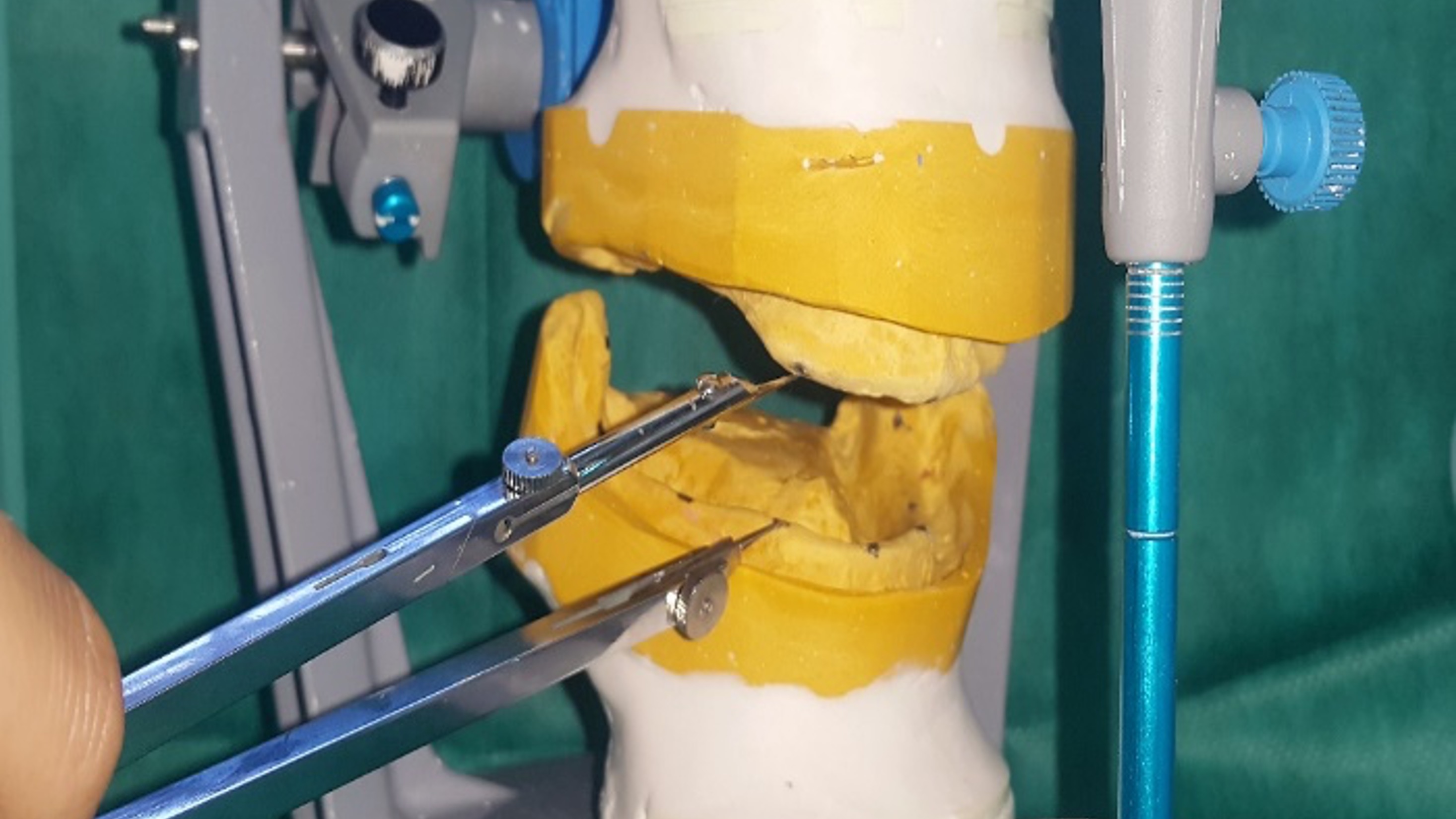
Méthodes : Une étude transversale a été menée sur 50 patients complètement édentés consultant pour une réhabilitation bimaxillaire à l'Hôpital Ibn Sina, Rabat. Après avoir enregistré lerapportmaxillo-mandibulaire, les modèles secondaires ont été montés sur articulateur pour mesurer la hauteur inter-crête, les dimensions des crêtes, et les périmètres des arcades. La résorption maxillaire et mandibulaire a été classifiée selon la classification de Cawood et Howell. Les données ont été analysées avec Jamovi 2.6.13.
Résultats : La hauteur inter-crête était plus grande au niveau médian [2 cm (1,7–2,2 cm) à droite ; 1,95 cm (1,7–2,3 cm) à gauche] et plus basse au niveau de la tubérosité-trigone [0,95 cm (0,7–1,48 cm) à droite ; 1,05 cm (0,8–1,48 cm) à gauche] (p < 0,001). La résorption maxillaire était surtout de classe III, tandis que la mandibule présentait principalement des classes IV et V, aussi bien en antérieur qu’en postérieur (p < 0,001).L'âge était significativement associé à la résorption mandibulaire antérieure (p < 0,05), et la durée de l'édentement était corrélée à la résorption dans les deux arcades (p < 0,05).....( résumé tronqué à 250 mots)
Téléchargements
Références
Ahuja S, Cagna DR. Defining available restorative space for implant overdentures. J Prosthet Dent.2010;104(2):133-6.
Heath MR. Facteurs cliniques qui déterminent l’espace prothétique. Congrès ADF 1972.Inf Dent. 1973;1:70-1.
Klein P. Quelques définitions pour mieux comprendre la piézographie.Inf Dent.1989; 71(29):2519-23.
Nabid A. Traité odontologique de piézologie. Alger: ENAG Editions; 2014. 592 p.
Kordatzis K, Wright PS, Meijer HJA. Posterior mandibular residual ridge resorption in patients with conventional dentures and implant overdentures. Int J Oral Maxillofac Implants. 2003;18(3):447-52.
Hirai T, Ishijima T, Hashikawa Y, Yajima T. Osteoporosis and reduction of residual ridge in edentulous patients. J Prosthet Dent. 1993;69(1):49-56.
Atwood DA. Some clinical factors related to rate of resorption of residual ridges. J Prosthet Dent. 2001;86(2):119-25.
Atwood DA. Bone loss of edentulous alveolar ridges. J Periodontol. 1979;50(4 Spec No):11-21.
Soikkonen K, Ainamo A, Xie Q. Height of the residual ridge and radiographic appearance of bony structure in the jaws of clinically edentulous elderly people. J Oral Rehabil. 1996;23(7):470-5.
de Baat C, Kalk W, van ’t Hof M. Factors connected with alveolar bone resorption among institutionalized elderly people. Community Dent Oral Epidemiol. 1993;21(5):317-20.
Nishimura I, Hosokawa R, Atwood DA. The knife-edge tendency in mandibular residual ridges in women. J Prosthet Dent. 1992;67(6):82-6.
Xie Q, Ainamo A, Tilvis R. Association of residual ridge resorption with systemic factors in home-living elderly subjects. Acta Odontol Scand. 1997;55(5):299-305.
Frankenthal S, Nakhoul F, Machtei EE, Green J, Ardekian L, Laufer D, et al. The effect of secondary hyperparathyroidism and hemodialysis therapy on alveolar bone and periodontium. J Clin Periodontol. 2002;29(6):479-83.
Imirzalioglu P, Yuzugullu B, Gulsahi A. Correlation between residual ridge resorption and radiomorphometric indices. Gerodontology. 2012;29(2):e536-42.
Bairam LR, Miller WA. Mandible bone resorption as determined from panoramic radiographs in edentulous male individuals ages 25-80 years. Gerodontology. 1994;11(2):80-5.
AlSheikh HA, AlZain S, Warsy A, AlMukaynizi F, AlThomali A. Mandibular residual ridge height in relation to age, gender and duration of edentulism in a Saudi population: A clinical and radiographic study. Saudi Dent J. 2019;31(2):258-264.
Parr NM, Passalacqua NV, Skorpinski K. Investigations into Age‐related Changes in the Human Mandible. J Forensic Sci. 2017;62(6):1586-1591.
Bressan E, Ferrarese N, Pramstraller M, Lops D, Farina R, Tomasi C. Ridge Dimensions of the Edentulous Mandible in Posterior Sextants: An Observational Study on Cone Beam Computed Tomography Radiographs. Implant Dent. 2017;26(1):66-72.
Cawood JI, Howell RA. A classification of the edentulous jaws. Int J Oral Maxillofac Surg. 1988;17(4):232-6.
Alidema SH, Bundevska J, Bimbashi V, Maja S, Dimoski G, Halili R. Cone Beam Computed Tomography (CBCT) Evaluation of Bone Height and Width in Edentulous Mandible. Int J Biomed. 2022;12(3):417-422.
Pietrokovski J, Starinsky R, Arensburg B, Kaffe I. Morphologic Characteristics of Bony Edentulous Jaws. J Prosthodont. 2007;16(2):141-7.
Pramstraller M, Schincaglia GP, Vecchiatini R, Farina R, Trombelli L. Alveolar ridge dimensions in mandibular posterior regions: a retrospective comparative study of dentate and edentulous sites using computerized tomography data. Surg Radiol Anat. 2018;40(12):1419-1428.
Huumonen S, Haikola B, Oikarinen K, Söderholm AL, Remes-Lyly T, Sipilä K. Residual ridge resorption, lower denture stability and subjective complaints among edentulous individuals. J Oral Rehabil. 2012;39(5):384-90.
Tayachi Y, Tayari O, Bendhief R, Jaouadi J. Bone Resorption Evaluation According to Measurements’ Panoramic Radiographs: A Tunisian Cross-Sectional Study. Saudi J Oral Dent Res. 2021;6(11):511-518.
Koç A, Erdem R. Evaluating Edentulous Mandible Alveolar Crest Dimensions in Posterior Region By Cone Beam Computed Tomography. East J Med. 2020;25(3):345-349.
Fichiers supplémentaires
Publiée
Numéro
Rubrique
Catégories
Licence
Merci de créditer les auteurs lors de toute citation : La Tunisie Médicale (2026)

Ce travail est disponible sous licence Creative Commons Attribution - Pas d'Utilisation Commerciale - Pas de Modification 4.0 International.
